When Its Just a Bump on the Head: Interpreting Skull Fractures in the Context of Child Maltreatment
Image: Sabra Snyder, SUNY Upstate Medical University, Medical Photography and Graphics
Accessible Version
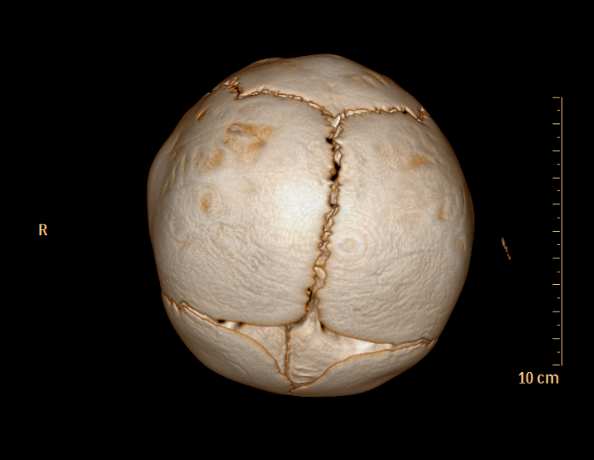
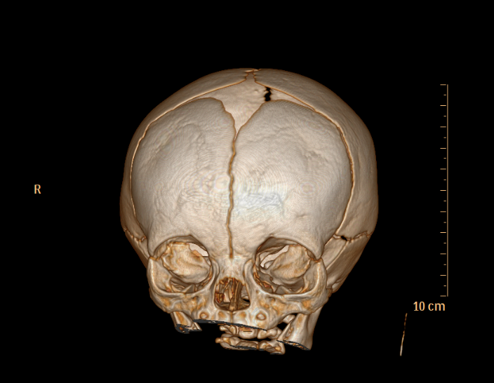
Let’s start with the normal anatomy of an infant skull. When I look at a CT scan, now more often than not a 3D reconstruction, I look at the normal structures paying close attention to the following: Are the sutures normal appearing and well approximated or is there significant diastasis to the sutures that might indicate increased intracranial pressure? Are there any accessory sutures that may be confused for a fracture Are there excessive (more than 10) Wormian bones – may be a hint of an underlying bone disease (e.g. Osteogenesis Imperfecta) Is there suggestion of soft tissue swelling and if so, can you tell where it is (ie. subgaleal? cephalohematoma?) As 3-D CTs have become more frequent and the technology has become more advanced, the answer to these questions has become easier for even a non-radiologist to answer.